What is Endoscopy?
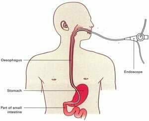
It is the direct visualization of a hollow organ or cavity inside our body by inserting a fiberoptic endoscope into it.
For example, the conventional gastroscope is a hollow, cylindrical metal tube that permits visualization of the inner surface of the stomach mucosa except for fundus, the greater curvature, and the pylorus.
Then introduced fiberscope, a type of gastroscope, has a shaft made for rubber or plastic that allows for greater curvature, the antrum, the pylorus, and the duodenal bulb.
Glass fibers that are incorporated into the shaft of the instrument transmit light to the mucosa and the image back to the examiner. A camera may be attached to either instrument for the purpose of taking pictures of abnormalities of the gastric mucosa during the examination.
What are some endoscopic procedures?
There are several types of endoscopy. The most common types of endoscopy for making a diagnosis but some are doing for surgical repair or correction. Also, some endoscopy is doing under direct vision Eg otoscopy, fundoscopy.
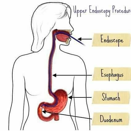
Those using natural body openings include esophagogastroduodenoscopy (EGD) which is often called upper endoscopy, gastroscopy, enteroscopy, endoscopic ultrasound (EUS), endoscopic retrograde cholangiopancreatography (ERCP), colonoscopy, and sigmoidoscopy
Some physicians recommend that a sigmoidoscopy be included in the annual physical examination of men over 40 because of the relatively high incidence of cancer of the rectum and sigmoid in this age-group.
Various Endoscopic Procedures and Their Purposes
| Types of Endoscopy | Purpose |
| Arthroscopy | Visualization of joint, usually knee and shoulder |
| Bronchoscopy | Visualization of the tracheobronchial tree |
| Choledochoscopy | Visualization of common bile ducts |
| Colonoscopy | Visualization of the colon |
| Culdoscopy | Visualization of pelvic organs |
| Cystoscopy | Visualization of the urinary bladder |
| Duodenoscopy | Visualization of the duodenum |
| Fundoscopy | Visualization of the fundus of the eye |
| Gastroscopy | Visualization of the stomach |
| Hysteroscopy | Visualization of the uterus |
| Jejunoscopy | Visualization of jejunum |
| Laparoscopy | Visualization of the abdominal organs |
| Laryngoscopy | Visualization of the larynx |
| Nephroscopy | Visualization of the renal pelvis |
| Esophagoscopy | Visualization of the esophagus |
| Ophthalmoscopy | Visualization of the fundus of the eye |
| Otoscopy | Visualization of the auditory canal and tympanic membrane |
| Peritoneoscopy | Visualization of the abdominal and pelvic organs within the peritoneal cavity |
| Pleuroscopy | Visualization of the pleural cavity |
| Proctoscopy | Visualization of the rectum and anal canal |
| Salpingoscopy | Visualization of the fallopian tube |
| Sigmoidoscopy | Visualization of the sigmoid colon and rectum |
| Thoracoscopy | Visualization of pleural surface in the thoracic cavity |
| Urethroscopy | Visualization of the urethra |
| Vascular endoscopy | Visualization of the lumen of blood vessels |
Some Common Questions About Endoscopy
How painful is endoscopy?
The patient is given sedatives 30 to 60 minutes before the examination or at the time of examination to lessen apprehension and decrease awareness of the passage of the instrument. An endoscopy isn’t typically agonizing, however, it tends to be awkward.
The vast majority just have gentle distress, like acid reflux or an irritated throat. The method is normally done while you’re alert. You might be given a local sedative to numb a particular part of your body.
To decrease secretions, atropine sulfate may be administered
Do they put you to bed for an endoscopy?
Every single endoscopic methodology includes some level of sedation, which loosens up you and curbs your gag reflex. A few times a mix of local sedative and gentle sedation is utilized to assuage the uneasiness.
Being calmed during the method will place you into a moderate to profound rest, so you won’t feel any inconvenience when the endoscope is embedded through the mouth and into the stomach.
To what extent does it take to recover from an endoscopy?
An upper endoscopy takes roughly 10 to 15 minutes. A colonoscopy takes around 15 to 30 minutes. Patients stay in the recovery zone 30 to 40 minutes after the procedure. The patient is instructed after the procedure not to eat or drink until the gag reflex returns, the least fluid aspirated into the lungs.
The gag reflux usually returns after 4 hours. A patient who goes home should check by touching the throat with a swab or finger before attempting to eat or drink
What is the preparation for an endoscopy?
Endoscopic Procedures of Upper G.I. Tract
Day of endoscopy
Nothing to eat or drink in any event 8 hours before the methodology. Medicine can be taken 4 hours before assessment with little taste of water. So that the patient does not regurgitate when the tubular instrument is passed through the mouth into the esophagus, and so that the lining of the stomach is visible. Try not to TAKE ANY ANTACIDS. Wear-free open dressing.
Occasionally an esophagoscopy or a gastroscopy must be performed as an emergency measure to remove a foreign object such as a bone or a pin. In such an emergency the stomach cannot be emptied, but suction should be available for use to prevent aspiration of regurgitated food or fluid.
Eyeglasses or dentures to be removed to prevent them are being broken. The patient should be void before the examination to prevent discomfort.
Endoscopic Procedures of Lower G.I. Endoscopy
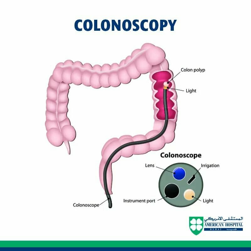
On the morning of the examination, enemas may be given until the return is clear, or a small hypertonic salt solution enema may be ordered.
If the patient is suspected of having ulcerative colitis, the physician will not probably order hypertonic enema, since it will cause inflammation of the mucosa and confuse the findings of the examination. Cathartic rectal suppositories like bisacodyl(Dulcolax) are also used.
Visualization of the bowel mucosa is impossible unless all the fecal material is evacuated.
Endoscopic Procedures of the Urinary System
Cystoscopy
Cystoscopy is the examination of the inside of the bladder through an instrument called a cystoscope. The cystoscope is connected to an illuminating source, thus enabling direct visualization of the bladder wall.
This procedure is indicated for patients who have or who have had hematuria.
Culdoscopy
A culdoscopy is an examination of the organs of female pelvis by the use of a culdoscope through the posterior fornix into the cul-de-sac of Douglas.
Peritoneoscopy and Laparoscopy
These procedures are similar procedures through which pelvic structures may be observed by approaching through the abdomen. Air may be injected and a radiograph was taken to outline pelvic structures fully