Table of Contents
What is Circle of Willis or Circulus Arteriosus?
Circle of Willis Anatomy

Circle of Willis is a ring of blood vessels at the base of the brain.
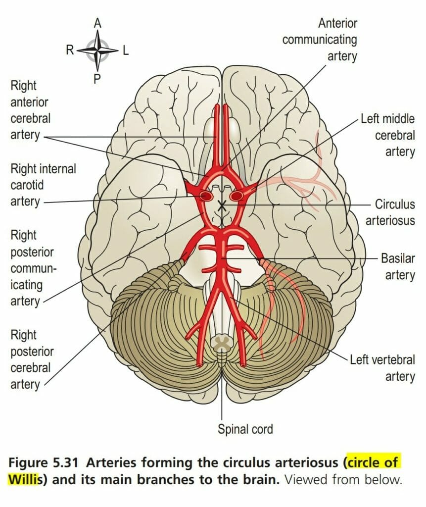
The greater part of the brain is supplied with arterial blood by an arrangement of arteries called Circle of Willis or Circulus arteriosus. The arrangement in the circle of Willis or Circulus arteriosus is such that the brain as a whole receives an adequate blood supply even when a contributing artery is damaged and during extreme movements of head and neck
The brain receives 20 percent of the cardiac output and uses 20 percent of body oxygen. Glucose is catabolized or burned for its energy. Blood flow is regulated by the levels of metabolite carbon dioxide. An increase in neuronal metabolism produces an increase in carbon dioxide, leading to local vasodilation. Local regulation of blood flow ensures that blood flow is proportional to neuronal metabolic needs.
Arteries of Circle of Willis
Four large arteries contribute to its formation, they are two internal carotid arteries (Left and Right) from both sides two vertebral arteries(Left and Right) from posterior sides
Anteriorly, the two anterior cerebral arteries arise from the internal carotid arteries and are joined by the anterior communicating artery
Posteriorly, the two vertebral arteries join to form the basilar artery. After traveling for a short distance the basilar artery divides to form two posterior cerebral arteries, each of which is joined to the corresponding internal carotid artery by a posterior communicating artery, completing the circle. The vertebral arteries are located on the anterolateral surface of the medulla

From this circle, the anterior cerebral arteries pass forward to supply the anterior part of the brain, the middle cerebral arteries pass laterally to supply the sides of the brain, and the posterior cerebral arteries supply the posterior part of the brain
Branches of the basilar artery supply parts of the brain stem.
The circle of Willis is formed by
- 2 anterior cerebral arteries
- 2 internal carotid arteries
- 1 anterior communicating artery
- 2 posterior communicating arteries
- 2 posterior cerebral arteries
- 1 basilar artery
Internal Carotid Artery
Internal Carotid artery supplies the greater part of the brain. It also has branches that supply eyes, forehead, and nose. It ascends to the base of the skull and passes through the carotid foramen in the temporal bone
Vertebral Artery
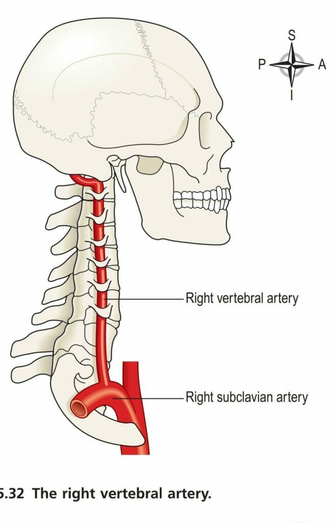
The vertebral arteries arise from the subclavian arteries pass upwards through the foramina in the transverse processes of the cervical vertebrae, enter the skull through the foramen magnum, then join to the basilar artery. The basilar artery bifurcates at the midbrain level to form two posterior cerebral arteries. The vertebral artery system supplies the brain stem, the cerebellum, the lower portion of the diencephalon, and the medial and inferior regions of the temporal and occipital lobes.
Venous Return

Venous blood from the head and neck is returned by deep and superficial veins.
Superficial veins with the same names as the branches of external carotid artery return venous blood from the superficial structures of the face and scalp and unite to form the external jugular vein.
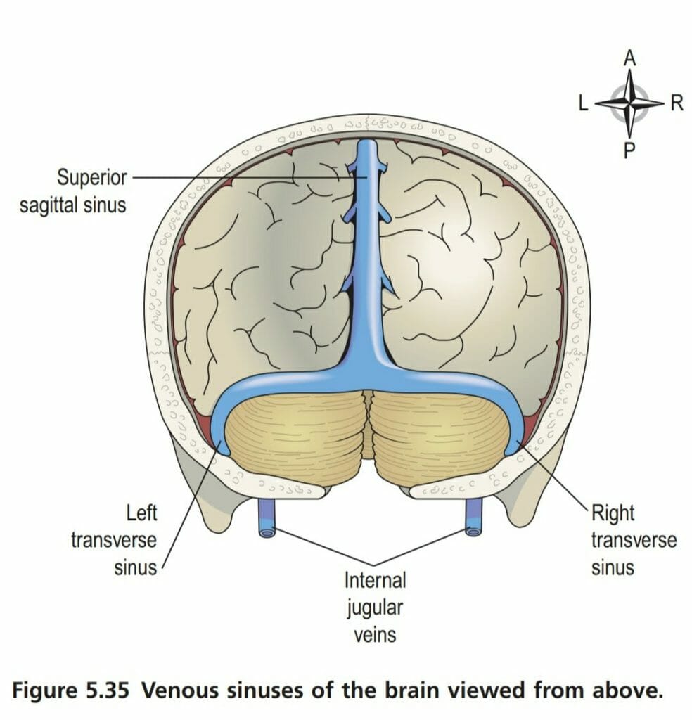
The venous blood from the deep areas of the brain is collected into channels called the dural venous sinuses, which are formed by layers of dura mater lined with endothelium.
The Main Venous Sinuses are Listed Below
- The superior sagittal sinus
- The inferior sagittal sinus
- The straight sinus
- The transverse sinuses
- The sigmoid sinuses